Magnetic Resonance Imaging

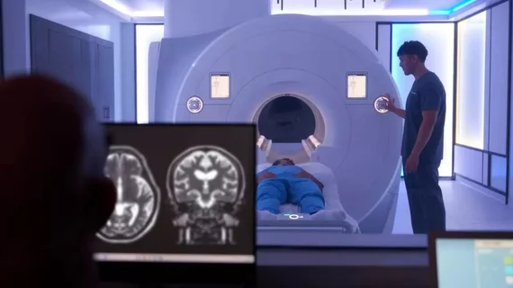
Magnetic resonance imaging (MRI) is the gold standard imaging modality for soft tissues. It produces detail cross-sectional images of soft tissue and bone anatomy, including muscles, tendons, ligaments, brain and organs, without the use of ionizing radiation. In addition to orthopedic imaging, MRI is also used for heart, brain and breast. MRI uses gadolinium contrast in many exams to highlight tissues and blood vessels, which enhances images and offers better diagnostic quality. It can also be used in conjunction with PET scans. How does MRI work? MR creates images by using powerful magnets to polarize hydrogen atoms in water (the body is made of of more than 80% water) so they face in one direction. A radiofrequency pulse is then used to ping these atoms, causing them to wobble, or resonate. The MRI coils detect this and computers can assemble images from the signals. Basic MRI scans will focus on the resonance of fat and water in two different sequences, which highlight and contrast different features in the anatomy.

Is that gin you're drinking? Nuclear MR spectroscopy may be able to tell you

Over the centuries, "gin" has become hard to define and even harder to classify due to its chemical diversity.

Thumbnail

UC San Diego earns $6.7M NIH grant to develop noninvasive imaging to quantify immune cells in tumors

The TAM-Sense technique is currently in pre-clinical development, but it could be used to develop new treatments.

Brains are getting bigger—and risk of dementia could be declining

MRI data from the Framingham Heart Study shows an increase in brain volume and surface area over a 40-year timeframe.

Thumbnail

Q&A: MRI accidents are rare, preventable—but safety standardization and certification are key

Emanuel Kanal, MD, founded the American Board of MR Safety to help develop safety standards for MR practices and to certify practitioners.

Thumbnail

Fire at VA hospital in Boston leads to closure of MRI suite

No one was injured in the incident, which originated from under the MRI machine.

SIR 2024: Study finds ultrasound ablation to be a precise, effective treatment for prostate cancer

The MRI-guided transurethral ultrasound ablation comes with minimal side effects, offering cancer patients an alternative to radiation or surgery.

Auburn University SIemens 7T magnetom scanner

University charts a world’s first with clinical use of new $9M 7T MRI system

The Siemens Magnetom scanner utilizes dedicated radiofrequency sodium coils and parallel transmit technology—a “stark” difference from older models

Thumbnail

Deep learning-based MRI reconstruction software produces considerable cost savings

Oulu University Hospital believes AI will allow it to deliver the same level of service with one fewer scanner, enabling annual savings of over $436,000.