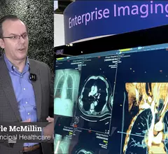
Enterprise Imaging

Enterprise imaging brings together all imaging exams, patient data and reports from across a healthcare system into one location to aid efficiency and economy of scale for data storage. This enables immediate access to images and reports any clinical user of the electronic medical record (EMR) across a healthcare system, regardless of location. Enterprise imaging (EI) systems replace the former system of using a variety of disparate, siloed picture archiving and communication systems (PACS), radiology information systems (RIS), and a variety of separate, dedicated workstations and logins to view or post-process different imaging modalities. Often these siloed systems cannot interoperate and cannot easily be connected. Web-based EI systems are becoming the standard across most healthcare systems to incorporate not only radiology, but also cardiology (CVIS), pathology and dozens of other departments to centralize all patient data into one cloud-based data storage and data management system.

Sectra to implement enterprise imaging SaaS in the cloud with Parkview Health in the US

International medical imaging IT and cybersecurity company Sectra (STO: SECT B) will implement its enterprise imaging cloud subscription service, Sectra One Cloud, at Parkview Health in the US. This will allow the Indiana-based health system future scalability as imaging volumes grow and will ensure data security in a fully managed cloud environment.

Sectra to implement enterprise imaging SaaS in the cloud with Parkview Health in the US

International medical imaging IT and cybersecurity company Sectra (STO: SECT B) will implement its enterprise imaging cloud subscription service, Sectra One Cloud, at Parkview Health in the US. This will allow the Indiana-based health system future scalability as imaging volumes grow and will ensure data security in a fully managed cloud environment.

Thumbnail

'Fictitious references' and 'significant inaccuracies' could hinder ChatGPT's medical writing career

Despite being widely praised by many of its users, ChatGPT recently left much to be desired by experts who compared the chatbot’s medical writing alongside that of seasoned professionals.

Thumbnail

AI model identifies radiologist-recommended follow-up imaging in reports, has potential for widespread use

New data published in the American Journal of Roentgenology details the performance of a deep learning model known as BERT, short for Bidirectional Encoder Representations from Transformers.

Decreasing energy consumption in radiology: How one hospital reduced use and saved big

Energy consumption reduction tactics could decrease greenhouse gas emissions owed to radiology while also saving departments tens of thousands of dollars every year. 

Thumbnail

EHR a key contributing factor to diagnostic errors in radiology, legal claims analysis finds

About 20% of the small sample of EHR-related errors occurred in radiology, informatics experts wrote in JAMA Network Open

radiology reporting EHR health record CDS AUC

Lessons learned from 7 years of structured radiology reporting at 1 institution

The University Medical Center Mainz recently surveyed radiologists and referrers to gather feedback on the change. 

Example of the four types of breast tissue density. The density of fibroglandular tissue inside the breast impacts the ability to easily see cancers. Cancers are very easy to spot in fatty breasts, but are almost impossible to find in extremely dense breasts. These examples show craniocaudal mammogram findings characterized as almost entirely fatty (far left), scattered areas of fibroglandular density (second from left), heterogeneously dense (second from right), and extremely dense (far right). RSNA

Experts developed a deep learning model that can estimate breast density

When tested, the model achieved a performance comparable to that of human experts.