Imaging Informatics

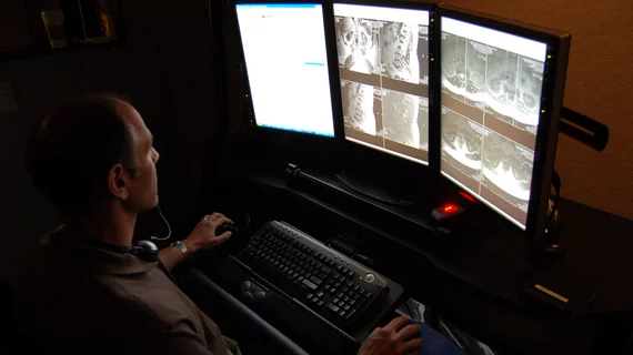
Imaging informatics (also known as radiology informatics, a component of wider medical or healthcare informatics) includes systems to transfer images and radiology data between radiologists, referring physicians, patients and the entire enterprise. This includes picture archiving and communication systems (PACS), wider enterprise image systems, radiology information. systems (RIS), connections to share data with the electronic medical record (EMR), and software to enable advanced visualization, reporting, artificial intelligence (AI) applications, analytics, exam ordering, clinical decision support, dictation, and remote image sharing and viewing systems.

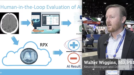
Closing the loop and settling rare clinical disagreements between radiologists at the same institution

Oftentimes, these deviations can occur informally, but experts believe careful documentation is needed to resolve them in a timely fashion.

Rankings of radiology IT solutions by end-users in the 2023 Best in KLAS program

End-users of various radiology IT systems offer their assessment of the software in the annual KLAS Research 2023 Best in KLAS report.

KLAS 2023 rankings for cardiovascular information systems and hemodynamic solutions

Hospital end-users ranked the CVIS and hemodynamic systems they are using and shed light on their working relationships with IT vendors. 

The integration of artificial intelligence (AI) into radiology PACS and enterprise imaging systems has become a big topic of discussion with IT vendors over the past couple years. This has become a bigger question from hospitals and radiology groups as there are now about 400 radiology related AI algorithms that have U.S. Food and Drug Administration (FDA) clearance. Amy Thompson, a senior analyst at Signify Research, is monitoring AI trends in radiology and discusses trends.

Trends in the adoption and integration of AI into radiology workflows

Amy Thompson, a senior analyst at Signify Research, explains why AI adoption has been slow in radiology, common barriers and trends in the market.

CT of coronavirus pneumonia, a solitary rounded ground-glass opacity (GGO) pattern. A 51-year-old woman in China presented in January 2020 without fever, but had close contact with positive patients. Top, baseline axial unenhanced chest CT obtained 6 days before the first positive PCR test. Bottom, chest CT scan 4 days later shows the size increase of the lesion (arrow). Image courtesy of RSNA. #COVID #SARSCoV2

How effective are chest CT severity scores in managing COVID?

While the studies on these systems have proven them to be effective in diagnosing and treating COVID in specific cohorts, the varying settings in which they were used can make it difficult to derive definitive conclusions on their efficacy.

Amy Thompson, a senior analyst at Signify Research, explains what she is seeing in the market for radiology PACS. She said the biggest overall, strategic technology trends are wider adoption of enterprise imaging systems expanding beyond radiology to include other departments, migration to cloud data storage, and adoption of artificial intelligence. Components of these integrate into the 5 trends in radiology IT systems outlined below.

5 key trends in PACS and enterprise imaging from Signify Research

Signify Research explains several key trends in the evolution of radiology PACS and enterprise imaging systems, including adoption of artificial intelligence, streamlining workflow, implementing structured reporting and more.

long covid lung CT

Some long COVID patients continue to display multi-organ damage one year after recovery

A new study utilizing multi-organ MRI scans recently identified organ impairment in 62% of COVID long haulers six months after their initial diagnosis; 29% of these individuals continued to display damage in at least one organ at the 12-month mark.

Thumbnail

Radiomics can predict major cardiac events using CCTA images

A CCTA-based radiomics method was recently found to be more accurate in identifying potentially problematic plaques than conventional CCTA anatomical parameters alone.