Pulmonary embolism

Pulmonary embolism (PE) is the third leading cardiovascular cause of death after heart attacks and stroke. PE is caused by blood clots in the pulmonary arteries. These are often caused by clots from the venous system, including thrombus from trauma, surgery or deep vein thrombosis (DVT). Treatment has traditionally been systemic use of thrombolytic drugs to dissolve the clot. But in cases there is a massive, life-threatening PE, or chronic clot burden that have remained in a vessel for an extended period of time, mechanical thrombectomy and ultrasound-assisted catheter-directed thrombolysis (USCDT) is being used as more targeted and aggressive treatments.

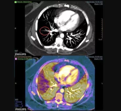
Example of pulmonary embolism (PE) detection on CT and confirmed using spectral CT. Image courtesy of Philips Healthcare

Researchers 'concerned' CT angiography topped MRA in follow-ups for pulmonary embolism

A recent study found whether a patient received MR angiography (MRA) instead of CT angiography (CTA) for a pulmonary embolism (PE) had little effect on the rate of follow-up chest CT or MRI one year later.

Thumbnail

American Airlines sued after passenger dies after in-flight pulmonary embolism

The family of a newlywed nurse is suing American Airlines after she fell ill on a flight and died. The cause of her death was determined to be an acute massive pulmonary embolism and cardiogenic shock.

Thumbnail

Teaming Up to Treat Pulmonary Emboli: Clotbusters Illustrates Potential of Coordinated Response Programs

At one hospital, an aggressive treatment PE treatment program has dramatically reduced mortality.

Additional MR Techniques Improve Detection of Pulmonary Embolism

According a study published in the March issue of the journal Radiology, radiologists now have a comparable, non-ionizing option to CT for the detection of pulmonary embolism.