Carotid artery disease causes up to 30% of all strokes, but severely calcified lesions can make treatment quite complex. The hope is that IVL can be as successful in this area as it has been in the treatment of coronary and peripheral artery disease.
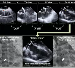
Heart teams can limit the risk of conduction disturbances that lead to permanent pacemaker implantation by utilizing both the cusp-overlap method and intracardiac echocardiography.
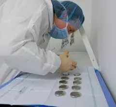
When patients with pacemakers die, what happens to the device? Typically, it ends up being discarded and forgotten—they were designed to be single-use devices, after all—but that does not have to be the case.


![Performing transcatheter aortic valve replacement (TAVR) with the J-Valve transcatheter heart valve (THV) may help limit the risk of coronary artery obstruction (CAO) in high-risk patients, according to a new study published in Clinical Interventions in Aging.[1]](/sites/default/files/styles/top_stories/public/2024-10/screenshot_2024-10-01_at_2.16.21_pm.png.webp?itok=kSiGReBW)