Cardiac Imaging

While cardiac ultrasound is the widely used imaging modality for heart assessments, computed tomography (CT), magnetic resonance imaging (MRI) and nuclear imaging are also used and are often complimentary, each offering specific details about the heart other modalities cannot. For this reason the clinical question being asked often determines the imaging test that will be used.

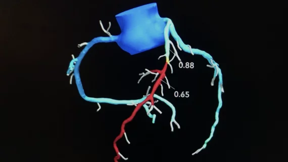
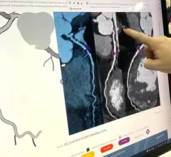
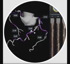
HeartFlow introduced its next generation artificial intelligence (AI) Plaque Analysis with an interactive experience at SCCT 2024. It shows a 3D plaque model and analysis by territory across calcified, non-calcified and low-attenuation plaques. This includes viewing cross-sectional, color-coded images of each plaque type where it was quantified along the vessel. #SCCT #SCCT24 #SCCT2024

Updated HeartFlow technology offers interactive features, full integration with FFR-CT 

HeartFlow is using SCCT 2024 to introduce the world to its updated Plaque Analysis platform. “Accurately diagnosing a patient’s risk for coronary artery disease is critical for determining the best treatment," Chief Medical Officer Campbell Rogers, MD, explained.

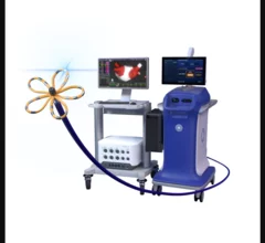
Cordis has received U.S. Food and Drug Administration (FDA) approval for its new MYNX Control Venous Vascular Closure Device (VCD) for 6F-12F access sites. The company hopes to launch the newly approved extravascular closure device in the months ahead.

Cordis gains FDA approval for new closure device, inks pricing deal with Premier

The newly approved extravascular closure device was designed for procedures with access sites from 6 to 12 French. It uses the same proprietary GRIP technology found in other Cordis interventional devices.

How the proposed Medicare Physician Fee Schedule could impact nuclear cardiology

The American Society of Nuclear Cardiology explored some key points included in the 2025 Medicare Physician Fee Schedule proposed rule.

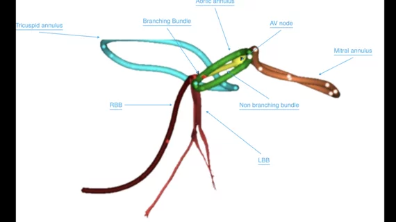
Researchers with University College London and the European Synchrotron Radiation Facility (ESRF) have used a new X-ray technique, hierarchical phase-contrast tomography (HiP-CT), to capture images of the human heart in unprecedented detail. The group shared its images, as well as a full analysis, in Radiology.[1]

New imaging technique captures human heart with 'unprecedented detail'

The new images were captured at the European Synchrotron Radiation Facility using hierarchical phase-contrast tomography. One specialist called them "Google Earth for the human heart." 

SCCT aims to provide common language for CCTA use with updated guidance

The Society of Cardiovascular Computed Tomography has developed a new expert consensus document designed to get physicians on the same page when discussing this growing technology. 

Emily Lau, MD, Mass General Hospital, discusses sex differences in cardiovascular presentations of women.

CVD presents differently in women than it does in men—guidelines should reflect that

Emily Lau, MD, a women's health expert with Massachusetts General Hospital, thinks major changes are needed in how we diagnose CVD in women. "Our guidelines need to be more precise and offer sex-specific recommendations," she says.

Video interview with Jim Melton, DO, vascular surgeon, explains the CLI mortality rate among amputees in rural Oklahoma, and the need for outreach programs to screen more patients.

Death rate from critical limb ischemia is high in rural Oklahoma

In rural Oklahoma, double amputees due to untreated peripheral artery disease face mortality rates around 35% after one year and 60% after two. Jim Melton, DO, hopes his mobile cardiology clinics can make a difference for these patients.

Healthcare salaries and compensation

Compensation keeps climbing in cardiology, electrophysiology, heart surgery

Among general cardiologists, compensation increased nearly 8% from 2023 to 2024. Interventional cardiology and electrophysiology saw even larger jumps.