Cardiac Imaging

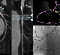
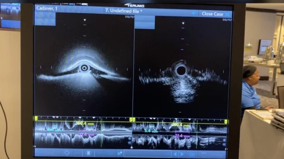
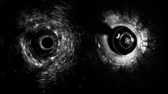
While cardiac ultrasound is the widely used imaging modality for heart assessments, computed tomography (CT), magnetic resonance imaging (MRI) and nuclear imaging are also used and are often complimentary, each offering specific details about the heart other modalities cannot. For this reason the clinical question being asked often determines the imaging test that will be used.

P‐waves can help identify Black patients prone to developing AFib

Research that followed patients for more than 13 years suggests increases in ECG PR interval, P‐wave duration and PTFV1 are associated with increased AFib risk in this cohort.

American Society of Nuclear Cardiology (ASNC) President-elect Jamieson M. Bourque, MD, MHS, FASNC, medical director of nuclear cardiology, echocardiography, and the stress laboratory at UVA Health in Charlottesville, and a professor of medicine and radiology at the University of Virginia, explains some of the new things ASNC will be doing in 2026

Collaborations, guidelines and grants: ASNC shares big plans for 2026

American Society of Nuclear Cardiology President-elect Jamieson M. Bourque, MD, detailed just some of what the group has planned for 2026. For example, ASNC will be working on the development of multiple new guidelines designed to help clinicians provide high-quality care.

Heartflow PCI Navigator Cardiology

New Heartflow tech delivers AI-powered PCI planning for cardiologists

PCI Navigator from Heartflow was designed to help cardiologists plan ahead for percutaneous coronary interventions before they even step foot into the cath lab. It assesses a patient’s CCTA results and creates a state-of-the-art 3D model.

PET study targets inflammation to predict AMI recovery

Inflammation-targeting PET method predicts recovery after heart attack

Targeting CXCR4 during PET scans could help providers gain vital information regarding patients' potential to fully recover from myocardial infarction. 

ASE leading push to revise CPT code for interventional echocardiography

The pressure is on for ASE and other societies to create a new CPT code for the use of interventional echocardiography during structural heart procedures.

Nuclear cardiology is entering a new era—one that goes well beyond the traditional focus on myocardial perfusion imaging. According to Marcelo Di Carli, MD, editor-in-chief of the Journal of Nuclear Cardiology and chief of the Division of Nuclear Medicine at Brigham and Women’s Hospital, the field is rapidly expanding into diagnosing and monitoring complex cardiac diseases such as amyloidosis, sarcoidosis, myocarditis, and cardiac infections.

Nuclear cardiology expands beyond perfusion imaging as it enters a new era

Beyond amyloidosis, nuclear cardiology is also increasingly used to image inflammation and infection in the myocardium, especially in diseases such as cardiac sarcoidosis and myocarditis.

Daniel Addison, MD, Director of Translational Research in the Cardiology Division and Associate Director for Survivorship and Outcomes Research in the Harold C. Simmons Comprehensive Cancer Center. He also holds the Audre and Bernard Rapport Endowed Chair in Cardiovascular Research, UT Southwestern Medical Center. explains the health disparities and inequities involved in cardio-oncology.

How clinicians can combat health disparities in cardio-oncology

Researchers, for example, could improve disparities by performing their tests on more diverse groups of patients. Daniel Addison, MD, shared his perspective with Cardiovascular Business. 

Positron emission tomography (PET) nuclear imaging has seen rapid expansion in cardiology and oncology due to its improve accuracy over SPECT and its ability to expand into new areas of cardiovascular imaging. A new review in the American Heart Association journal Stroke outlines another possible use of PET to assess carotid artery atherosclerosis for stroke prevention. Figure courtesy Stroke, https://www.ahajournals.org/doi/10.1161/STROKEAHA.125.050399

PET holds promise for carotid plaque assessments

PET has already gained significant momentum in recent years. Now, researchers are pointing to another possible use for the popular imaging modality: assessing carotid artery atherosclerosis.