Cardiac Imaging

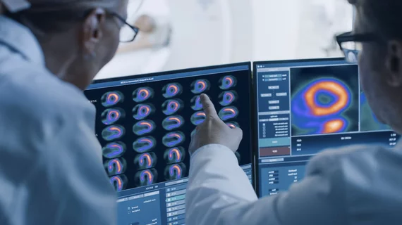
While cardiac ultrasound is the widely used imaging modality for heart assessments, computed tomography (CT), magnetic resonance imaging (MRI) and nuclear imaging are also used and are often complimentary, each offering specific details about the heart other modalities cannot. For this reason the clinical question being asked often determines the imaging test that will be used.

An Illinois appellate judge upheld a pervious trial victory that cleared and central Illinois cardiologist Amit Dande, MD, and Prairie Cardiovascular Consults LLP of alleged misdiagnosis of the severity of a patient's heart condition that led to his death prior to a schedule percutaneous coronary intervention (PCI).

Illinois cardiologist not held responsible for patient's death while waiting for angiogram

A judge upheld a previous trial victory that cleared Prairie Cardiovascular Consults of alleged mishandling of a patient and not fully understanding the severity of the heart condition prior to their death.

Examples of photon-counting coronary angiography showing how clarity improves as the thickness of the image is reduced. Top: 60-year-old female, with noncalcified plaque (arrowheads) and coronary stenosis (inset images). The reduced section thickness did not affect assessment in this patient. Bottom: 56-year-old female with calcified plaque (arrowheads) and coronary stenosis. The reduced section thickness leads to less calcium blooming and therefore a less severe percentage of stenosis. Courtesy of RSNA

Coronary artery disease classification improved with photon-counting CT

After a photon-counting CT, 54% of patients had their coronary artery disease classification downgraded.

Examples of the messages the Nanox AI algorithms display for incidental findings of spinal compression fractures and detection of coronary calcium. Both can help physicians better understand risk factors or need for therapy in patients through these types of opportunistic screenings on scans being performed for other reasons.

Routine chest CT often reveals patients at risk for cardiovascular disease, presenting radiology with ‘untapped’ potential

Rads only reported this incidental finding in about 31% to 44% of cases, experts detailed in the Journal of the American College of Radiology

Luc Mertens, MD, section head of echocardiography at The Hospital for Sick Children in Toronto, explains application of ultrafast ultrasound and blood speckle imaging in echocardiography

Vector flow and blood speckle echocardiography imaging may unveil new clinical insights

Luc Mertens, MD, Hospital for Sick Children, Toronto, explains research and potential uses for ultrafast echo imaging.

Thumbnail

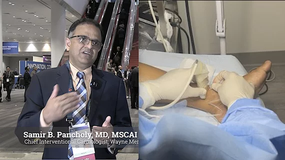
Implementing a same-day PCI discharge pathway boils down to correct patient selection

Safety is key to which individuals are selected for SDD, and there can be benefits to both patients and health systems as this becomes the predominant model of care in elective PCI.

Staff at the new Fetal Cardiac Clinic at the Heart Institute at Children's Hospital Los Angeles. From left, Director of the Heart Institute David Romberger, RN, MSN, CCRN; attending physician Shuo Wang, MD; Executive Director of the Heart Institute Jennifer Klunder, MHA; Director of the Fetal Cardiac Clinic Jodie Votava-Smith, MD; Chief of the Division of Cardiology and Co-director of the Heart Institute Paul F. Kantor, MBBCh, MSc, FRCPC; and congenital cardiac surgeon Luke Wiggins, MD.

Children’s Hospital Los Angeles unveils new fetal cardiac clinic

Clinic for expectant mothers designed to better diagnose and treat congenital heart defects and heart diseases before birth. 

An example of cardiac MRI myocardial blood flow reserve (MBFR) imaging. Image courtesy of https://doi.org/10.3390/diagnostics10090679

Imaging group thrilled with new CPT codes for MBFR measurements

The Society for Cardiovascular Magnetic Resonance applauded two new CPT Category III codes for myocardial blood flow reserve (MBFR) assessments.

Michael Garshick, MD, director of the cardio-rheumatology program, cardiologist and assistant professor, Ronald O. Perelman Department of Dermatology, NYU Grossman School of Medicine, explains the role of inflammation in coronary disease development and how this risk factor can be detected and treated.

Targeting coronary inflammation helps cardiologists provide better care

Michael Garshick, MD, examines the role of inflammation in coronary disease development, detailing how this risk factor can be detected and treated.