Cardiac Imaging

While cardiac ultrasound is the widely used imaging modality for heart assessments, computed tomography (CT), magnetic resonance imaging (MRI) and nuclear imaging are also used and are often complimentary, each offering specific details about the heart other modalities cannot. For this reason the clinical question being asked often determines the imaging test that will be used.

Thumbnail

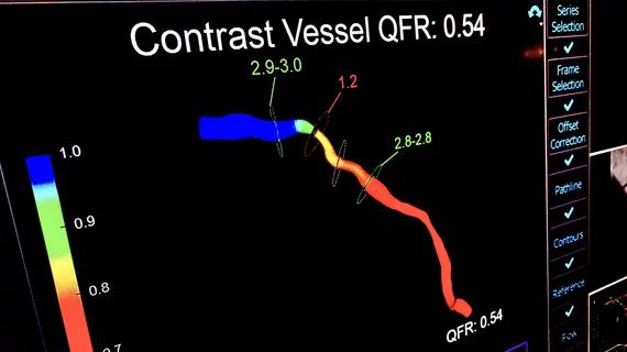
Angiography-derived FFR accurately diagnoses normal, abnormal vessel function

Fractional flow reserve (FFR) can be closely approximated using conventional coronary angiography and propriety software, according to the FAST-FFR study published online in Circulation. The findings offer a potential route for more patients with suspected coronary artery disease to receive functional assessment of lesions without the need for a guidewire or hyperemic agents.

Thumbnail

Novel imaging approach tracks atherosclerotic plaque buildup in vivo

Researchers at the University of Tsukuba in Japan have identified a new approach for imaging the progression of atherosclerotic plaque in vivo, according to a study published in Scientific Reports.

Thumbnail

Axial chest CT IDs chamber enlargement with high specificity, reasonable sensitivity

Cardiac chamber enlargement can be identified with high specificity and reasonable sensitivity on axial chest CT images by use of gender-specific measurement thresholds, according to new research published in the American Journal of Roentgenology.

Carestream Digital X-ray System Deployed at Remote Antarctic Station for Diagnosis of Illnesses, Injuries

Scientific Station Staffed All Year Despite Nine Months Of Temperatures That Can Dip to -120 Degrees Fahrenheit

Thumbnail

CT-based measurements beat Framingham risk score in predicting future CVD events

Assessment of abdominal aortic calcification using computed tomography (CT) is a highly effective predictor of future heart attack and CVD risk, according to research published in Radiology Oct. 2—more effective even than the Framingham risk score, which has been relied upon for more than two decades.

Thumbnail

CT reveals abdominal aortic calcification can predict future heart attack, stroke

Heart attack and other life-threatening cardiovascular events can be predicted with CT-based measures of calcification in the abdominal aorta and could be a more accurate predictive model than the widely known Framingham risk score, according to research published Oct. 2 in Radiology.

Thumbnail

Deep learning, SPECT-MPI forecast obstructive CAD

SPECT myocardial perfusion imaging (MPI) and deep learning have proven to be a powerful pair in predicting obstructive coronary artery disease (CAD) and improving the interpretation of upright and supine images, according to research published Sept. 27 in the Journal of Nuclear Medicine.

Thumbnail

How much is too much radiation in medical imaging?

As imaging scans before widely more available and affordable, more Americans are getting more tests than they used to—currently totaling to more than 85 million CT scans each year, according to an article published Oct. 1 by The Washington Post. But how much is too much radiation?