Cardiac Imaging

While cardiac ultrasound is the widely used imaging modality for heart assessments, computed tomography (CT), magnetic resonance imaging (MRI) and nuclear imaging are also used and are often complimentary, each offering specific details about the heart other modalities cannot. For this reason the clinical question being asked often determines the imaging test that will be used.

Polio-like condition detected with MRI is targeting US children

A polio-like condition called acute flaccid mylelitis (AFM) that causes partial paralysis is showing up in children across the U.S. and can only be detected with MRI and spinal fluid tests, according to a report published Oct. 8 by NBC News.

Thumbnail

Carotid artery MRI boosts risk assessment for CVD, stroke

MRI measurements of carotid wall thickness improve predictions of cardiovascular disease risk more than a commonly used ultrasound-based test, according to research published Oct. 9 in Radiology.

FDA Clears MAGNETOM Sola 1.5T MRI Scanner From Siemens Healthineers

The Food and Drug Administration (FDA) has cleared the MAGNETOM Sola, a 1.5 Tesla magnetic resonance imaging (MRI) scanner from Siemens Healthineers that brings groundbreaking BioMatrix technology to the 1.5T market.

Thumbnail

Carotid artery MRI improves CVD, stroke risk assessment

Measuring wall thickness in carotid arteries with MRI may help better determine cardiovascular disease risk assessment, according to research published Oct. 9 in Radiology.

Canon Medical Launches Healthy Sonographer Program

First-of-Its-Kind Program Aims to Help Prevent Sonographer Injuries While Enhancing Patient Care and Reducing Costs.

Thumbnail

ACR appoints 1st female chief research officer, Etta Pisano

The American College of Radiology (ACR) has named breast imaging researcher Etta Pisano, MD, chief research officer of the ACR, the first woman in ACR's history to hold the position.

Thumbnail

Angiography-derived FFR accurately diagnoses normal, abnormal vessel function

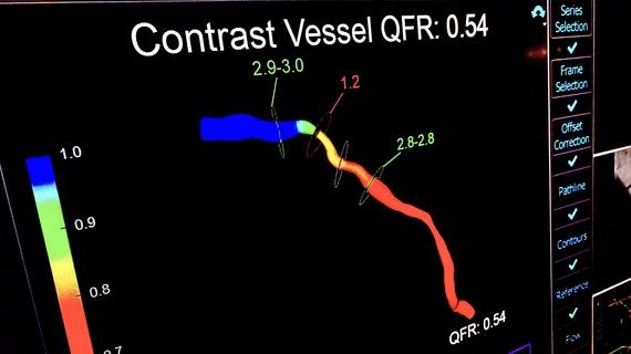
Fractional flow reserve (FFR) can be closely approximated using conventional coronary angiography and propriety software, according to the FAST-FFR study published online in Circulation. The findings offer a potential route for more patients with suspected coronary artery disease to receive functional assessment of lesions without the need for a guidewire or hyperemic agents.

Thumbnail

Novel imaging approach tracks atherosclerotic plaque buildup in vivo

Researchers at the University of Tsukuba in Japan have identified a new approach for imaging the progression of atherosclerotic plaque in vivo, according to a study published in Scientific Reports.