Cardiac Imaging

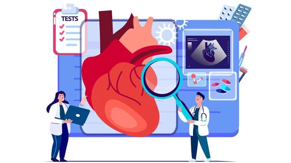
While cardiac ultrasound is the widely used imaging modality for heart assessments, computed tomography (CT), magnetic resonance imaging (MRI) and nuclear imaging are also used and are often complimentary, each offering specific details about the heart other modalities cannot. For this reason the clinical question being asked often determines the imaging test that will be used.

Thumbnail

UK radiology crisis left 56K angina patients without lifesaving scans in 2017

Critical shortages of imaging equipment and qualified radiologists left an estimated 56,000 angina patients without access to potentially lifesaving computed tomography (CT) scans last year, the Royal College of Radiologists and British Society of Cardiovascular Imaging announced Nov. 8.

Thumbnail

56K angina patients in UK denied CTs in 2017 due to radiologist shortage

Last year, more than 56,000 angina patients across the U.K. were denied life-saving CT heart scans due to the continuing shortage of radiologists and scanners, according to data released Nov. 8 by the Royal College of Radiologists (RCR) and the British Society of Cardiovascular Imaging (BSCI), published in The BMJ.

The Eluvia drug-eluting stent continues to demonstrate positive outcomes in long lesion IMPERIAL sub-study

Boston Scientific announced clinical outcomes from the IMPERIAL Long Lesion Sub-study demonstrating that the Eluvia Drug-Eluting Vascular Stent System is safe and effective in treating patients with long, complex, calcified lesions within the superficial femoral and proximal popliteal arteries.

‘Everyone will feel the pinch’: Nuclear imaging labs brace for Tc-99m shortage

Nuclear medicine clinics could be operating at 25 percent capacity or less by the end of the week due to a shortage of Technetium-99m, said one hospital leader.

Thumbnail

CAC scoring may ID patients best suited for statins

Coronary artery calcium (CAC) scoring could be the key to determining which patients are most likely to benefit from statin therapy, according to a retrospective analysis published Nov. 5 in the Journal of the American College of Cardiology.

Thumbnail

Automated valve tracking reduces time, variability in 4D flow MRI

Automated cardiac valve tracking halved the analysis time and variability in blood-flow quantification with four-dimensional (4D) flow MRI, researchers reported in Radiology. The algorithm may help clinicians more efficiently assess the severity of congenital and valvular heart disease, they said.

Thumbnail

4D flow MRI may improve automated valve tracking in congenital heart patients

Researchers from the Netherlands demonstrated, in a study published Oct. 30 in Radiology, that four-dimensional (4D) flow MRI with automated cardiac valve tracking reduces analysis time and improves reliability of valvular flow quantification compared to manual valve tracking in patients with congenital heart disease.

Thumbnail

Augmented reality allows 3D visualization of myocardial scar during ablation

An augmented reality (AR) technique involving 3D visualization of myocardial scar can assist physicians in evaluating and treating cardiac damage during electrophysiological interventions like ventricular tachycardia (VT) ablation, researchers reported in PLOS One.