Cardiac Imaging

While cardiac ultrasound is the widely used imaging modality for heart assessments, computed tomography (CT), magnetic resonance imaging (MRI) and nuclear imaging are also used and are often complimentary, each offering specific details about the heart other modalities cannot. For this reason the clinical question being asked often determines the imaging test that will be used.

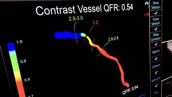
Left, HeartFlow's RoadMap analysis enables cardiac CT readers to identify stenoses in the major coronary arteries. The AI provides visualization and quantification of the location and severity of anatomic narrowings. Right image, HeartFlow's Plaque Analysis AI algorithm automates assessment of coronary plaque characteristics and volume on CCTA exams to greatly reduce the time it takes to manually assess and quantify these features.

HeartFlow gains FDA clearance for 2 new AI-powered imaging assessments

The solutions, Plaque Analysis and RoadMap Analysis, both use coronary CT angiography to provide clinicians with a noninvasive look at patients who present with coronary artery disease and face a heightened myocardial infarction risk.

transcarotid TAVR TAVI transcatheter aortic valve replacement

Transcarotid TAVR a safe alternative when femoral access is not an option

The conventional transfemoral approach is not always possible during TAVR procedures, leaving care teams with an important decision to make. 

The Medtronic SelectSecure MRI SureScan Model 3830 cardiac lead already gained FDA approval for His-Bundle pacing back in 2018. Now, the lead is also approved for left bundle branch area pacing, providing users with another option when treating patients for bradycardia. According to Medtronic, this marks the first time the FDA has approved a device for this indication.

Medtronic cardiac lead the first to receive FDA approval for conduction system pacing

The company’s SelectSecure MRI SureScan Model 3830 cardiac lead already gained FDA approval for His-Bundle pacing back in 2018. Now, the lead is also approved for left bundle branch area pacing.

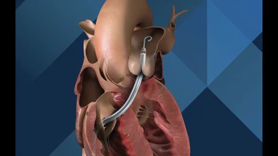
Impella Heart Pump Abiomed RECOVER IV RCT cardiogenic shock

Impella heart pumps linked to improved high-risk PCI outcomes

Researchers examined data from patients who underwent high-risk PCI with either an Impella heart pump or an intra-aortic balloon pump.

VIDEO: Cardiac CT as a front-line chest pain exam in the ED

Harold Litt, MD, explains the use of cardiac CT as a primary imaging exam for chest pain at his emergency department over the past decade.

An updated look at how cognitive impairment affects TAVR outcomes

The connection between cognitive impairment and postoperative delirium among TAVR patients stood out as a definite concern to the study's authors.

Thumbnail

FAPI PET/CT findings linked with risk of sudden cardiac death

Experts involved in the research suggested that these findings indicate a potential role for FAPI PET/CT imaging in detecting changes in myocardial fibrosis, noting that it could be more sensitive to earlier changes than standard cardiac MRI.

CT has been used for planning and procedural guidance in pulmonary vein isolation (PVI) for about 20 years. It shows the anatomy to the electrophysiologist. The EPs also can load the imaging into their EP mapping system. It also shows them the location of the esophagus and the phrenic nerve so they can avoid these during the procedures. #PVI #EPeeps

VIDEO: The expanding role of cardiac CT in electrophysiology

Harold Litt, MD, division chief of cardiothoracic imaging and director of the Center for Advanced CT Imaging Sciences at the Perelman School of Medicine of the University of Pennsylvania, explains the role of cardiac CT in EP procedures.