Nuclear Cardiology

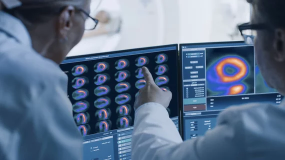
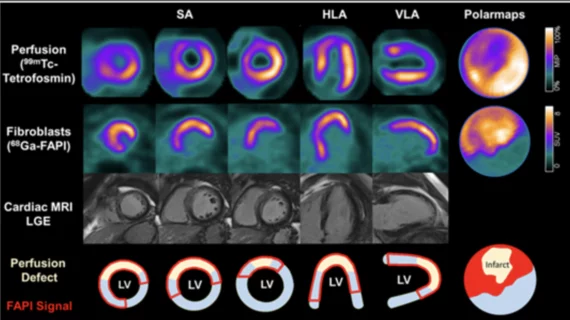
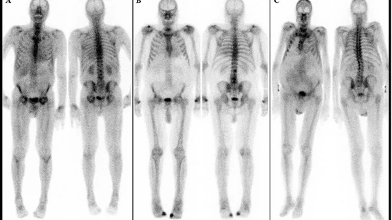
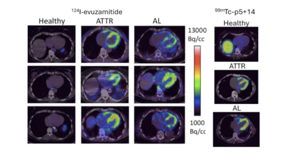
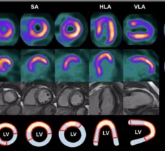
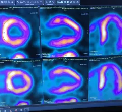
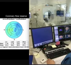
Single photon computed tomography (SPECT) and positron emission tomography (PET) molecular imaging are used as primary cardiac imaging modalities to evaluate the function of the heart. It uses radioactive isotopes attached to sugars that are metabolized by cardiomyocytes. This creates an image of the metabolic activity of the heart and shows areas of ischemia or infarct. Other radiotracers can image the heart to diagnosis cardiac amyloidosis and sarcoidosis. 

Thumbnail

SNMMI backs updated ASNC guidelines on SPECT MPI

New 2018 guidelines for myocardial perfusion imaging (MPI) protocols on conventional and novel single photon emission computed tomography (SPECT) for nuclear cardiology practitioners has received praise from the Society for Nuclear Medicine and Molecular Imaging (SNMMI).

Thumbnail

Cleveland Clinic's Cremer on cardiac imaging in cardiovascular oncology

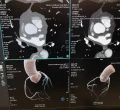
At this year's "Nuclear Cardiology Today: Best Practices for Today, Innovation for Tomorrow" conference in Rosemont, Illinois, sponsored by the American Society of Nuclear Cardiology (ASNC), Paul Cremer, MD, discussed the role of cardiac imaging in cardio-oncology.
Thumbnail

PET technique visualizes amyloid deposits in heart

PET with 11C-PIB provides a noninvasive method for visualizing amyloid deposits in the heart, according to a study published in the February issue of the Journal of Nuclear Medicine. The researchers suggest that 11C-PIB eventually may be used in the clinical setting as both a diagnostic tool and a treatment follow-up method.

ASNC Releases Dose-Reduction Guidelines for Nuclear Cardiology

Technological improvements in image acquisition and software processing in nuclear cardiology should allow physicians to shave patient imaging times dramatically or cut radiation doses fourfold, according to a new preferred practice statement from the American Society of Nuclear Cardiology (ASNC).