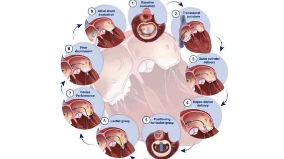
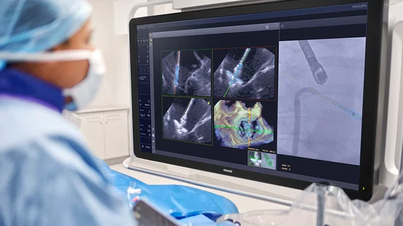
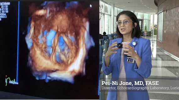
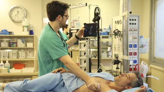
Echocardiography

Cardiac ultrasound uses reflected sound waves (echos) to create images of anatomy inside the body. Echocardiograms are the primary cardiac imaging modality used to assess the heart and diagnose or track cardiac issues. Echo is the gold standard imaging modality to assess the heart, particularly with calculating left ventricular ejection fraction (LVEF), which is a measure of cardiac output. In addition to noninvasive standard transthoracic echo (TTE), invasive transesophgeal echo (TEE) is also used when clearer, more detailed imaging of the heart is needed. Both 3D and 4D echo echo systems are rapidly gaining wider adoption and enable new types of assessments, especially in the structural heart space and in transcatheter procedural guidance. Find news on general ultrasound imaging.

William A. Zoghbi, MD, MACC, FAHA, FASE, is the chair of the Department of Cardiology at the Houston Methodist DeBakey Heart and Vascular Center, and past president of both the American College of Cardiology (ACC) and the American Society of Echocardiography (ASE).

VIDEO: New advances in echocardiography

William A. Zoghbi, MD, past president of the ACC and ASE, discussed the latest trends in cardiac ultrasound technology. 

March 15, 2022
The mitral valve seen on 3D cardiac ultrasound offers a noninvasive surgeon's view of the valve for better diagnostics and pre-planning of structural heart procedures. Example from the GE Vivid E95 FlexiLight photo-realistic 3D/4D lighting system.

Q&A: William Zoghbi breaks down the future of echocardiography

In a new interview, former ACC and ASE President William A. Zoghbi, MD, reviews some of the biggest trends in cardiac ultrasound.

February 24, 2022
Tiny fragments of plastic are commonly found deep inside the human body. Heart surgery, it seems, is one of many ways these microplastics are reaching their destination.  Surgeons Operating On Patient

TEE improves 30-day outcomes for patients undergoing cardiac valve or proximal aortic surgery

Intraoperative TEE, researchers wrote, can provide value during any open cardiac valve or proximal aortic surgery.

February 10, 2022
kort_stock.jpg

Seeking Out Severe Aortic Stenosis: The Low Down on Low Flow-Low Gradient

Sponsored by Medtronic

It’s not uncommon for severe aortic stenosis to go unrecognized, and thus untreated. When the data points to the existence of low-flow, low-gradient aortic stenosis, a diagnosis can be even more challenging.

February 7, 2022

Medtech company Us2.ai gains FDA clearance for automated echocardiography software

The news comes following a validation study at Brigham and Women's Hospital and multiple trials with pharmaceutical partners.

September 15, 2021
Even patients who were never hospitalized for COVID-19 face a heightened risk of cardiovascular issues. #longcovid

Imaging technique could help predict heart rhythm issues among COVID-19 patients

"This is a safe and affordable new data point that can clue us in about who might develop atrial fibrillation," one researcher said. 

July 12, 2021

Echocardiogram add-on predicts patients at risk of developing heart problems from COVID-19

Speckle-tracking strain is a safe, affordable way to gain clues into who may develop atrial fibrillation and require additional monitoring down the road.

July 9, 2021
Researchers recently completed an external validation for the Ovarian-Adnexal Reporting and Data System (O-RADS) for classifying lesions as malignant or benign, for which validation had been previously lacking. #ORADS

No practice changes needed after ultrasound-related FDA safety warning, imaging experts say

Such injectable agents have been administered routinely for years to detect heart disease, assess risk, and identify tumors throughout the abdomen.

May 12, 2021