Artificial Intelligence

Artificial intelligence (AI) is becoming a crucial component of healthcare to help augment physicians and make them more efficient. In medical imaging, it is helping radiologists more efficiently manage PACS worklists, enable structured reporting, auto detect injuries and diseases, and to pull in relevant prior exams and patient data. In cardiology, AI is helping automate tasks and measurements on imaging and in reporting systems, guides novice echo users to improve imaging and accuracy, and can risk stratify patients. AI includes deep learning algorithms, machine learning, computer-aided detection (CAD) systems, and convolutional neural networks. 

neck ultrasound thyroid

TI-RADS could help guide thyroid nodule biopsy decisions in children

ACR TI-RADS has previously been shown to lack sensitivity for distinguishing between benign and malignant thyroid lesions in pediatric patients.

MRI radiomics could change the future of breast cancer treatment

Radiomics methodologies could change how care plans are managed for patients with breast cancer by identifying those most likely to benefit from specific treatments.

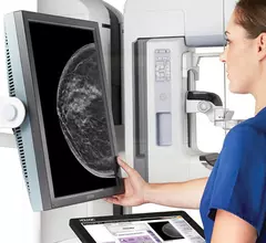
An example of commercially available artificial intelligence (AI) automated grading of breast density on mammograms from the vendor Densitas..

VIDEO: Role of AI in breast imaging with radiomics, detection of breast density and lesions

Connie Lehman, MD, chief of breast imaging, co-director of the Avon Comprehensive Breast Evaluation Center at Massachusetts General Hospital, discusses how artificial intelligence (AI) is being implemented in breast imaging.

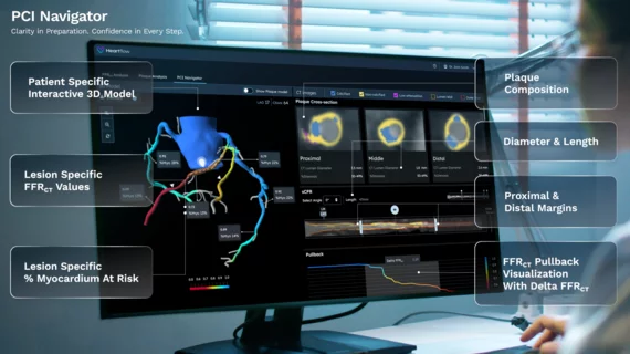
Left, HeartFlow's RoadMap analysis enables cardiac CT readers to identify stenoses in the major coronary arteries. The AI provides visualization and quantification of the location and severity of anatomic narrowings. Right image, HeartFlow's Plaque Analysis AI algorithm automates assessment of coronary plaque characteristics and volume on CCTA exams to greatly reduce the time it takes to manually assess and quantify these features.

HeartFlow gains FDA clearance for 2 new AI-powered imaging assessments

The solutions, Plaque Analysis and RoadMap Analysis, both use coronary CT angiography to provide clinicians with a noninvasive look at patients who present with coronary artery disease and face a heightened myocardial infarction risk.

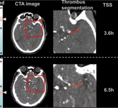
AI system boosts intracranial hemorrhage detection

“This study implies that future clinical workflows may see AI be used in an adjunct capacity to improve interpretations of CT scans by helping call radiologists' attention to findings that may be overlooked.” 

Example of a cancer that is difficult to see in dense breast tissue, but can be seen easier using 3D mammography digital breast tomosynthesis (DBT) breast imaging because the radiologist can go through the breast layer by layer if tissue..

VIDEO: The rapid adoption of 3D mammography and use of AI to address dense breasts

Stamatia Destounis, MD, a radiologist and managing partner at Elizabeth Wende Breast Care in Rochester, New York, chair of the American College of Radiology (ACR) Breast Commission, explains the rapid adoption of 3D mammogram digital breast tomosynthesis (DBT) technology.
 

Society of Breast Imaging (SBI) President John Lewin, MD, explains some of new initiatives and technology in mammography to increase earlier breast cancer detection. #SBI #breastimaging #mammography

VIDEO: SBI president outlines trends in breast imaging

Society of Breast Imaging President John Lewin, MD, explains some of the new initiatives and technology in mammography that are designed to increase early breast cancer detection.

pacs.jpg

VIDEO: KLAS shares trends in enterprise imaging and AI

Monique Rasband, vice president of imaging, cardiology and oncology, KLAS Research, explains some of technology trends KLAS researchers have found in enterprise imaging system and radiology artificial intelligence (AI).