Computed Tomography

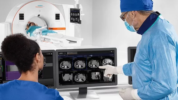
Computed tomography (CT) is a fast and accurate imaging modality often used in emergency settings and trauma imaging. CT scans, with or without (or both) iodinated contrast are frequently used to image the brain, chest, abdomen and pelvis, but also have post-imaging reconstructive capabilities for detailed orthopedic imaging. It is now a standard imaging modality in emergency rooms to quickly assess patients. CT uses a series of X-ray images shot as the gantry rotates around the patient. Computer technology assembles these into into a dataset volume than can be slices on any access, or advanced visualization software can extract specific parts of the anatomy for study. Find more content specific to cardiac CT.

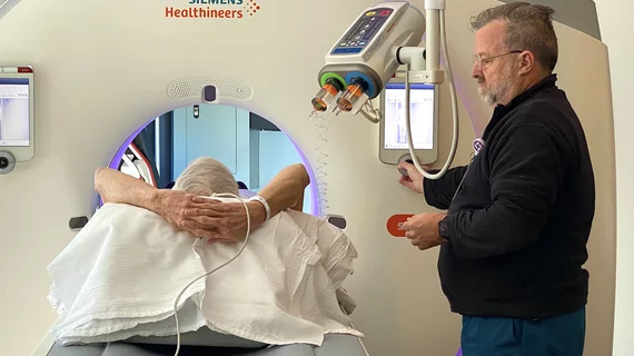
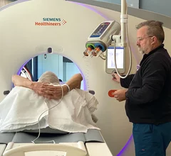
The imaging iodine contrast shortage is delaying procedures and causing rationing at hospitals. impact is it having on hospitals and the tough decisions that are being made to triage patients to determine if they will get a contrast CT scan or an interventional or surgical procedure requiring contrast. Photo by Dave Fornell

Experts developing AI model that learns from calcium-scoring CT scans

The team hopes to develop a model that will estimate a patient's risk of major cardiovascular events and predict when such events are most likely to occur.

Standard contrast protocols may be 'overdosing' women with the imaging agent

New research suggests female patients attenuate significantly more contrast media in their vessels and organs compared to men. 

The imaging iodine contrast shortage is delaying procedures and causing rationing at hospitals. impact is it having on hospitals and the tough decisions that are being made to triage patients to determine if they will get a contrast CT scan or an interventional or surgical procedure requiring contrast. Photo by Dave Fornell

Multidose contrast injectors could increase waste in some radiology departments

These findings are important from both environmental and operational standpoints, authors of a new Academic Radiology paper point out. 

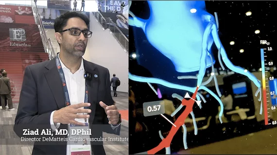
Video of Deepak Bhatt explaining details on the TRANSFORM trial using CCTA and AI. #TCT #TCT24 #TCT2024 #AHA24 #AHA2024

New cardiac prevention paradigm explored in TRANSFORM trial using AI and CCTA

Deepak Bhatt, MD, director of the Mount Sinai Fuster Heart Hospital and principal investigator of the TRANSFORM trial, explains an emerging technique for cardiac screening: combining coronary CT angiography with artificial intelligence for plaque analysis to create an approach similar to mammography.

PHOTO GALLERY: Medical imaging technology at RSNA 2024

Take a virtual tour around the Radiological Society of North America (RSNA) meeting to see the sights and new technologies displayed across the vast exhibit hall floors.

Ischemic stroke shown in CT scans. Image courtesy of RSNA

New algorithm is twice as accurate at predicting stroke timing compared to the standard of care

Determining stroke onset is critical for management, as there is a small window of time for initiating treatment that can inhibit damage.

New protocol addresses contrast allergies in patients with suspected stroke

In patients with suspected stroke, contrast allergies present a significant dilemma, as contrast-enhanced imaging is often used to guide treatment decisions.

lung cancer pulmonary nodule chest

Radiology Partners study unearths 3 common barriers to lung cancer screening

RP affiliate Desert Radiology surveyed 300 screening-eligible patients during their visits, sharing the findings in the journal of Cancer Epidemiology