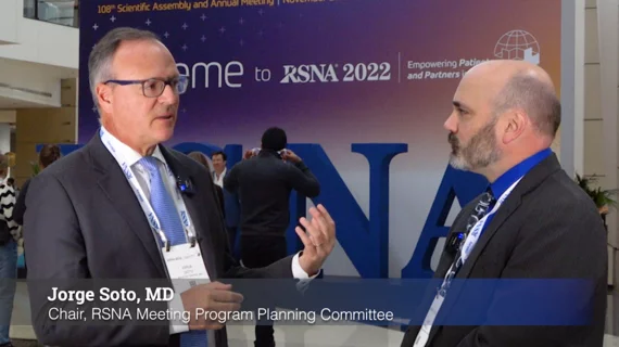
Lung scarring occurs in up to 11% of patients recovered from COVID, regardless of infection severity

Patient with severe, lung damage from COVID. Images in a 54-year-old man with COVID-19–related acute respiratory distress syndrome and subsequent fibrosis. (A) Axial CT 2 weeks after admission shows diffuse ground-glass opacity (GGO) with reticular abnormality and traction bronchiectasis in right middle lobe, indicating organizing phase of lung injury. (B) Axial CT 6 months after admission shows decreased GGO but extensive traction bronchiectasis and architectural distortion, suggesting fibrosis. RSNA

Patient with severe, long-term lung damage from COVID. Images in a 54-year-old man with COVID-19–related acute respiratory distress syndrome and subsequent fibrosis. (A) Axial CT 2 weeks after admission shows diffuse ground-glass opacity (GGO) with reticular abnormality and traction bronchiectasis in right middle lobe, indicating organizing phase of lung injury. (B) Axial CT 6 months after admission shows decreased GGO but extensive traction bronchiectasis and architectural distortion, suggesting fibrosis. Patient remained symptomatic with restricted pulmonary function. Image courtesy of RSNA. Read more. Read more.

Researchers cannot yet determine whether these changes will progress or resolve in the long term.