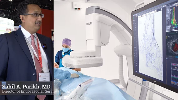
Sahil Parikh, MD, director of endovascular services at Columbia University Irving Medical Center, said treating these patients can be quite challenging.
Alexandra Bastiany, MD, made history when she became Canada’s first Black female interventional cardiologist in 2020. Now, that achievement is being recognized in a surprising way.
Reventics, a revenue cycle management company and a business associate of Regional One Health, detected a cyber-intruder who accessed the company’s servers in December 2022.
“It's all about risks and benefits," one researcher said. "Starting transitioning is a big part of a person's life and helping them feel more themselves, but hormone replacement therapy also has a lot of side effects—it's not a risk-free endeavor."
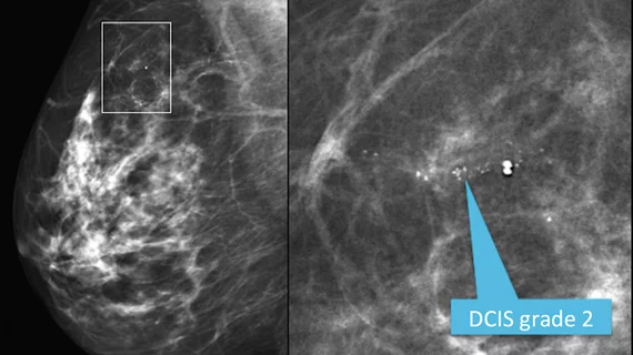
Mediolateral oblique view from a screening mammogram in a 54-year-old woman shows a small cluster of microcalcifications in the upper outer quadrant of the right breast. The right image shows a detailed spot magnification view of the calcifications. Stereotactic biopsy revealed grade 2 ductal carcinoma in situ (DCIS). See the next image for an MRI view. Read more. RSNA image.
New research suggests that the tissue environment where microcalcifications of the breast are formed could hold clues into how breast cancer progresses.
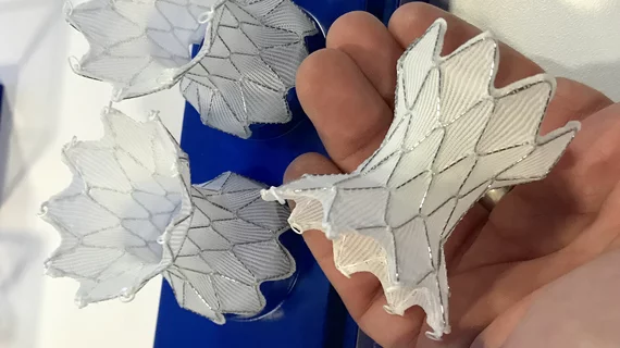
The device, which gained FDA approval in 2021, was part of a voluntary recall in 2022 due to stability concerns. Medtronic worked with the FDA to address the issue, and the Harmony TPV is once again available throughout the United States.