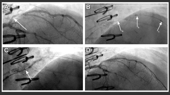
Angiography demonstrates a high-grade, modestly angulated stenosis (arrow) in the proximal segment of a large diagonal branch (A). Predilation performed with difficulty, and the GEC could not be advanced beyond stenosis even with “inch worming” techniques. (B). Advancement of the CrossFAST (C) beyond the proximal stent edge stenosis (a.), the position of the distal end of the outer catheter (b.), and the microcatheter tip (c.) are indicated. Photo and caption courtesy of JSCAI