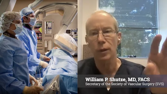
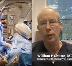
Vascular surgeons perform procedures interventional cardiologists and interventional radiologists do not do—so why not make sure they are part of every heart team conversation?
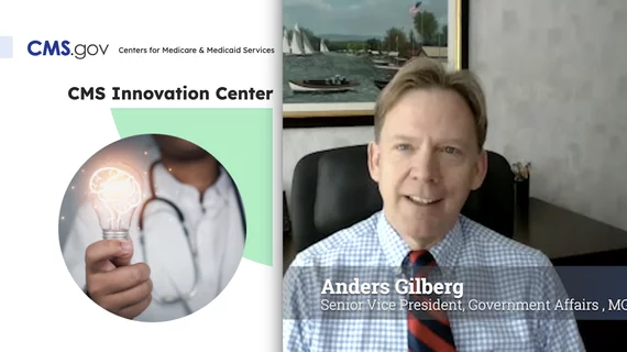
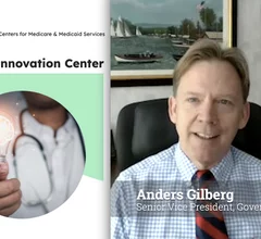
The Center for Medicare and Medicaid Innovation (CMMI) has tested nearly 40 APMs, of which only two turned out to be successful, which does not bode well for meeting a 2030 deadline to transition to value-based payments.
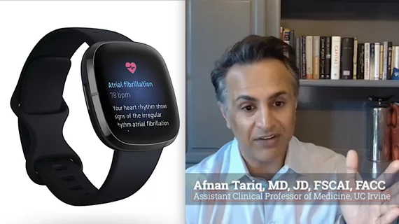
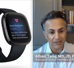
Afnan Tariq, MD, discusses early data on a passive, device-agnostic AI platform for heart failure monitoring. “When clinicians are empowered with insights and able to act earlier, you're able to have a durable impact," he said.
Vascular surgeons perform procedures interventional cardiologists and interventional radiologists do not do—so why not make sure they are part of every heart team conversation?
The Center for Medicare and Medicaid Innovation (CMMI) has tested nearly 40 APMs, of which only two turned out to be successful, which does not bode well for meeting a 2030 deadline to transition to value-based payments.
Afnan Tariq, MD, discusses early data on a passive, device-agnostic AI platform for heart failure monitoring. “When clinicians are empowered with insights and able to act earlier, you're able to have a durable impact," he said.
While the Trump administration is pushing to accelerate clinical AI adoption, SCAI has emphasized that physicians must guide how the technology is implemented and governed.
The Medical Group Management Association explains some positive federal healthcare policy movement since the start of 2026, including extending telehealth and rural health payments, and possible solutions to the prior authorization burden in Medicare Advantage.