Nuclear Cardiology

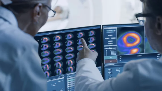
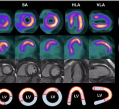
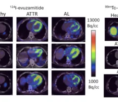
Single photon computed tomography (SPECT) and positron emission tomography (PET) molecular imaging are used as primary cardiac imaging modalities to evaluate the function of the heart. It uses radioactive isotopes attached to sugars that are metabolized by cardiomyocytes. This creates an image of the metabolic activity of the heart and shows areas of ischemia or infarct. Other radiotracers can image the heart to diagnosis cardiac amyloidosis and sarcoidosis. 

The new cardiac PET radiotracer flurpiridaz F-18 is posed to be a major game changer and will likely lead to increased adoption of cardiac PET.

Largest network of private cardiology groups in US to deploy GE HealthCare's cardiac PET tracer

One of the tracer’s more significant advantages is its extended half-life of 109 minutes—significantly longer than other currently available PET MPI imaging agents.

American Society of Nuclear Cardiology (ASNC) President-elect Jamieson M. Bourque, MD, MHS, FASNC, medical director of nuclear cardiology, echocardiography, and the stress laboratory at UVA Health in Charlottesville, and a professor of medicine and radiology at the University of Virginia, explains some of the new things ASNC will be doing in 2026

Collaborations, guidelines and grants: ASNC shares big plans for 2026

American Society of Nuclear Cardiology President-elect Jamieson M. Bourque, MD, detailed just some of what the group has planned for 2026. For example, ASNC will be working on the development of multiple new guidelines designed to help clinicians provide high-quality care.

PET study targets inflammation to predict AMI recovery

Inflammation-targeting PET method predicts recovery after heart attack

Targeting CXCR4 during PET scans could help providers gain vital information regarding patients' potential to fully recover from myocardial infarction. 

Nuclear cardiology is entering a new era—one that goes well beyond the traditional focus on myocardial perfusion imaging. According to Marcelo Di Carli, MD, editor-in-chief of the Journal of Nuclear Cardiology and chief of the Division of Nuclear Medicine at Brigham and Women’s Hospital, the field is rapidly expanding into diagnosing and monitoring complex cardiac diseases such as amyloidosis, sarcoidosis, myocarditis, and cardiac infections.

Nuclear cardiology expands beyond perfusion imaging as it enters a new era

Beyond amyloidosis, nuclear cardiology is also increasingly used to image inflammation and infection in the myocardium, especially in diseases such as cardiac sarcoidosis and myocarditis.

Positron emission tomography (PET) nuclear imaging has seen rapid expansion in cardiology and oncology due to its improve accuracy over SPECT and its ability to expand into new areas of cardiovascular imaging. A new review in the American Heart Association journal Stroke outlines another possible use of PET to assess carotid artery atherosclerosis for stroke prevention. Figure courtesy Stroke, https://www.ahajournals.org/doi/10.1161/STROKEAHA.125.050399

PET holds promise for carotid plaque assessments

PET has already gained significant momentum in recent years. Now, researchers are pointing to another possible use for the popular imaging modality: assessing carotid artery atherosclerosis.

Marcelo Di Carli, MD, editor-in-Chief of the Journal of Nuclear Cardiology, chief, Division of Nuclear Medicine and Molecular Imaging, Department of Radiology, and executive director, Cardiovascular Imaging Program, at Brigham and Women's Hospital, explains why there is rapid growth of PET imaging because of the extra value it brings in detecting microvascular disease.

How PET is transforming cardiac nuclear imaging

"PET has changed the practice of nuclear medicine in general and I think it is going to revitalize the practice of cardiology,"  Marcelo Di Carli, MD, told Cardiovascular Business. He noted that more practices seem interested in implementing PET than ever before.

American Society of Nuclear Cardiology (ASNC) President-elect Jamieson M. Bourque, MD, MHS, FASNC, medical director of nuclear cardiology, echocardiography, and the stress laboratory at UVA Health in Charlottesville, and a professor of medicine and radiology at the University of Virginia, explains key trends and his plans to advance nuclear cardiology in 2026. #ASNC

Incoming ASNC president making AI's integration into nuclear cardiology a top priority

Jamieson M. Bourque, MD, MHS, previewed his plans for the future in a new interview with Cardiovascular Business. 

Imagers reviewing flurpiridaz F-18 (Flyrcado) myocardial perfusion PET images. Image from GE Healthcare

Nuclear imaging groups share new flurpiridaz guideline

Several groups collaborated on the document, which is designed to help clinicians provide high-quality, consistent care when using flurpiridaz during cardiac PET imaging exams.