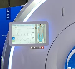
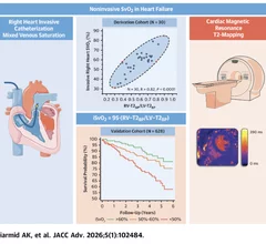
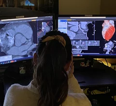
Magnetic Resonance Imaging

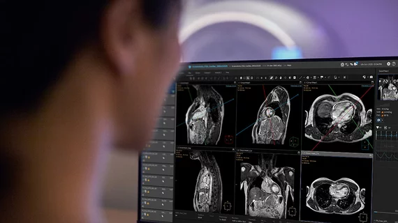
Cardiac magnetic resonance imaging MRI is used as both a functional and anatomical cardiac imaging test. It offers excellent soft tissue detail and the ability to quantify cardiac function. MRI scans can be performed with or without gadolinium contrast depending on what information is needed. Unlike computed tomography (CT), MRI does not use X-ray radiation, but patients with metal implants may have contraindications for MRI use because MR will heat up most metal objects. MRI exams usually take much longer than CT scans. How does MRI work? MR creates images by using powerful magnets to polarize hydrogen atoms in water (the body is made of of more than 80% water) so they face in one direction. A radiofrequency pulse is then used to ping these atoms, causing them to wobble, or resonate. The MRI coils detect this and computers can assemble images from the signals. Basic MRI scans will focus on the resonance of fat and water in two different sequences, which highlight and contrast different features in the anatomy.

artificial intelligence heart AI

FDA clears AI-powered cardiac imaging platform

The Tempus Pixel platform was designed to assist clinicians with the evaluation of cardiac MR images.

pericarditis cardiac imaging

Pericarditis: New guidance takes on the diagnosis and management of a challenging heart condition

Pericarditis, inflammation of the pericardium, accounts for approximately 5% of all emergency department evaluations for chest pain. These new recommendations are designed to help guide clinicians through the ins and outs of patient care. 

An example of artificial intelligence automated coronary artery calcium scoring shown by Coreline at ACC23. The AI also identifies and color codes the anatomy so help show the reviewing radiologist or cardiac imager what and where things were calculated. #ACC #ACC23

Cardiology still No. 2 in FDA-cleared clinical AI algorithms, trailing only radiology

Cardiology continues to be one of the most influential healthcare specialties when it comes to utilizing artificial intelligence. 

heart data AI algorithm cardiologist doctor

New AI model outperforms humans at predicting sudden cardiac death

The model spots previously undetected scarring patterns on cardiac MRI scans of patients with hypertrophic cardiomyopathy—a leading cause of sudden cardiac death among young people.

heart data research doctor cardiologist AI

AI turns low-quality MRI scans into helpful heart images

A team of biomedical engineers, radiologists and cardiologists has developed a new AI model capable of restoring low-quality cardiac MRI scans.

Thumbnail

Severe COVID causes long-lasting coronary microvascular dysfunction, new data show

New MRI data demonstrate that patients who weathered severe cases of COVID-19 may sustain long-lasting heart damage. 

UTA's lower body negative-pressure pants for MRI stress tests

Inspired by astronauts, researchers use high-tech pants to uncover heart issues on MRI

New first-in-human data highlight the potential of pants designed to improve the quality of MRI-based cardiac stress tests.

GE HealthCare SIGNA Sprint MRI

GE HealthCare debuts new wide-bore MRI scanner built with cardiology, oncology in mind

The new 1.5T MRI scanner includes a wide bore and key AI features designed to boost the patient experience.