Nuclear Medicine

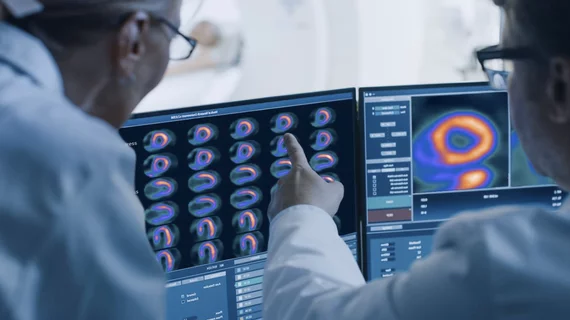
Nuclear medicine (also called molecular imaging) includes positron emission computed tomography (PET) and single photon emission computed tomography (SPECT) imaging. Nuclear imaging is achieved by injecting small amounts of radioactive material (radiopharmaceuticals) into patients before or during their scan. These can use sugars or chemical traits to bond to specific cells. The radioactive material is taken up by cells that consume the sugars. The radiation emitted from inside the body is detected by photon detectors outside the body. Computers take the data to assemble images of the radiation emissions. Nuclear images may appear fuzzy or ghostly rather than the sharper resolution from MRI and CT.  But, it provides metabolic information at a cellular level, showing if there are defects in the function of the heart, areas of very high metabolic activity associated with cancer cells, or areas of inflammation, data not available from other modalities. These noninvasive imaging exams are used to diagnose cancer, heart disease, Alzheimer’s and Parkinson’s disease, bone disorders and other disorders. 

Bill that would require providers report extravasations of radioactive drugs is back on the House floor

Patient advocacy groups say the proposal "is common-sense legislation that will protect patients."

Lantheus

Lantheus completes acquisition of radiopharma firm Evergreen Theragnostics for up to $1B

Founded in 2019 and headquartered in Springfield, New Jersey, Evergreen develops and manufactures imaging agents for diagnosing and treating cancer. 

GE Healthcare and Nihon Medi-Physics

GE HealthCare finalizes purchase of radiopharma firm that generates $183M in revenue

The Chicago-based manufacturer is buying the remaining stake in Nihon Medi-Physics, a Japanese developer of imaging agents used in SPECT and PET exams. 

PET method offers new insight into inflammation of the brain

Researchers are hopeful that their technique could improve the diagnosis of conditions associated with neural inflammation. 

Most popular radiology video interviews on Health Imaging. The evolving role of artificial intelligence (AI) in breast imaging was a big topic at the Radiological Society of North America (RSNA) 2024 meeting. Health Imaging spoke with Manisha Bahl, MD, breast imaging division quality director and breast imaging division co-service chief at Massachusetts General Hospital and an associate professor of radiology, Harvard Medical School, at the conference to hear more about her breast AI sessions.

Most popular radiology video interviews on Health Imaging

Radiology has seen a lot of large language model and generative AI research and adoption and it is clearly a hot button topic with our No. 1 video.

Thumbnail

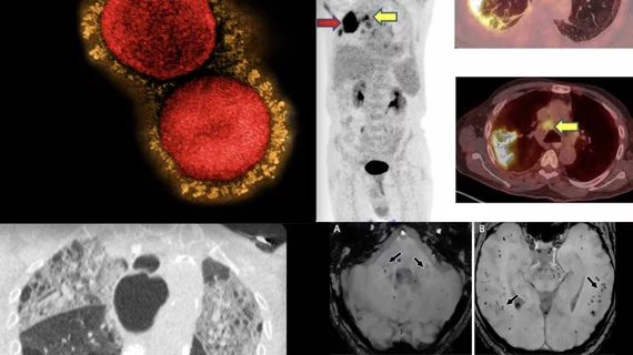
New PET imaging method could improve our understanding of long COVID

Researchers have developed a new radiotracer that targets one of the virus’ spike proteins, allowing them to track it longitudinally.  

FDA approves new prostate cancer imaging agent with extended shelf life

The new product from Telix paves the way for increased access at clinics where prostate PET may not have been available in the past.

Researchers track dopamine receptors to study its impact on cognitive function.

New PET study offers insight into how dopamine drives executive function

These findings could help guide the development of new treatments for psychiatric and neurological disorders.